Δευτέρα 24 Φεβρουαρίου 2014

Fibromyalgia

Fibromyalgia is a condition, described in medical physiology, as the presence of pain in the fibrous tissues and muscle (fibro- fibrous tissue; myo- muscle; algos- pain in Greek and Latin language) due to several reasons. This disease is primarily characterized by chronic pain and allodynia, which is characterized by extreme and heightened painful response to the tactile pressure.

Κυριακή 23 Φεβρουαρίου 2014

Knee Injuries in Basketball


Basketball is an exciting, fast-moving sport. Unfortunately, what makes the game exciting to watch and exhilarating to play also makes it a high-risk sport for knee injuries. Running with sudden stops, cutting side to side, jumping, and pivoting can injure your knee. Although there are risks of knee injury, it's still a great game, so before you jump in, you should know the risks and what you can do to prevent injury.

The largest joint in the body, the knee, is made up of the lower end of the femur (thighbone) and the upper end of the tibia, or shinbone. The patella, or kneecap, slides in a groove at the end of the femur. Ligaments at the end of the femur and tibia connect the bones and help stabilize and support the knee. Tendons connect the muscles to the bones, and the cartilage inside the joint helps to cushion and absorb shock to the joint and to give stability to the knee.

A sprain or strain can occur when there is a direct blow to the knee or when there is a sudden, stressful movement that affects the knee. A sprain or strain can also develop from overuse of the joint or when you place too much stress on the knee for a long period of time. Often, with a sprain or strain the tissues become irritated, causing pain and swelling.

Sprains
A sprain is a stretch or tear of a ligament. More serious sprains involve complete tears of one or more of the knee ligaments. A common knee sprain involves the anterior cruciate ligament (ACL). Changing direction rapidly or stopping abruptly while running can cause the twisting motion that tears the ACL.

If you sprain your knee, you may hear a popping or snapping sound at the time of injury. Afterward, pain seems to come from within the knee, especially with movement. You will not be able to bear weight on that leg, and you can experience swelling and fluid behind the kneecap. You may also hesitate to place weight on the knee because it feels loose or unstable.

Strains
Strains are defined as a partially or completely torn muscle or tendon. With knee strains, you may feel symptoms similar to a sprain and may see bruising around the injured area.

Patellar tendinitis, or jumper's knee, is a common strain that usually results from overuse. Jumper's knee is an inflammatory condition that causes pain in the front of the knee. The extensor mechanism, which includes the quadriceps muscle and patellar tendon, connects the patella (kneecap) to the femur (thighbone) and the tibia (shinbone) (Fig. 2). Patellar tendinitis begins as inflammation of the patellar tendon where it attaches to the patella. It can worsen by continual tearing or from degeneration of the tendon.

Treatment
You should rest your knee at the first sign of a sprain or strain. Immediately after injury, apply the RICE (rest, ice, compression and elevation) method. You should rest your knee as much as possible, apply ice packs for a couple of days to bring down the swelling, use compression, such as an ACE bandage, and elevate the leg on a pillow. For inflammation and pain, your doctor may prescribe anti-inflammatory medications such as aspirin or ibuprofen.

Treatment for knee injuries, such as severe sprains or strains (where a ligament or tendon is more seriously stretched or torn), may involve using a knee splint, immobilizer, or cast, and using crutches for a few weeks or months.

Depending on the injury, your doctor may suggest rehabilitation for your knee injury. Working with a physical therapist, you will do specific exercises designed to take your knee through its range of motion to prevent stiffness and scarring as your knee heals. You may also need to do regular exercises to strengthen the muscles surrounding the knee.

Injury prevention
To prevent knee injuries, always wear appropriate protective equipment during practices and competition. Kneepads and shinguards will help to protect your knee from injury. You'll also want to make sure you wear supportive shoes that are in good condition and are appropriate for the basketball court.

During workouts, always warm-up and cool down, and remember to increase your training intensity slowly. You might try weight lifting to strengthen your muscles and use stretching exercises to improve your flexibility because strong flexible muscles help support and protect joints. Warm-up with jumping jacks, a stationary bike, or running or walking in place for 3 to 5 minutes. Then slowly and gently stretch, holding each stretch for 30 seconds. After an intense workout, practice, or competition, slow down your heartbeat and stretch your muscles again to cool down. Do not suddenly increase the intensity or duration of your workout because it can lead to an overuse injury. If you play only one sport, maintain coordination and balance by training year-round even if it's at a lower intensity than during your competitive season.

The way you move can also help to prevent knee injuries. When you jump, bend your knees when you land to take pressure off the ACL. When you cut laterally or pivot, crouch at the hip and bend your knees to reduce your chance of ligament injury.

Never play through the pain of a sprain or strain. A minor injury can become a more serious injury that requires surgery if left untreated. A minor sprain or strain may keep you off the basketball court for a few weeks, but a major injury may keep you out of the game the entire season. Always see your doctor at the first sign of knee injury. You'll be glad you did.


What Is a Motor Unit?


A motor unit consists of one alpha motor neuron together with all the muscle fibers it stimulates. Since the human body contains, on average, 250,000,000 muscle cells and approximately 420,000 motor neurons, a motor unit will generally consist of a single motor neuron paired with many muscle fibers. In strength training, the early strength gains seen by novices are often not gains in size or number of muscle fibers, but activation of motor units that had been previously dormant.

The motor neuron is a specialized type of nervous cell that runs between the central nervous system and the muscles. Neurons typically consist of a cell body — the axon — and the dendrites. If a neuron were to be seen as a tree, the axon would be analogous to the trunk and the dendrites to the branches. Neurons found within the brain normally have relatively short axons, but neurons that are part of a motor unit — because they must connect to the muscles of the body — have elongated axons that run through the spinal cord, and out to the associated muscle fibers. Each muscle fiber is connected to a particular dendrite, and it is through the dendrites that messages are relayed between the central nervous system and the muscle fiber.

Muscle fibers are elongated cells, specialized to carry out the functions of the specific muscles of which they are a part. This is true of the cardiac muscles of the heart, the smooth muscles that make up the lining of many internal organs, and skeletal muscles. Only skeletal muscles, however, are under conscious control. The size and shape of the muscle fiber is dependent upon its function, with the smooth muscle cells being flattened and tile-like; skeletal muscle cells, long and rope-like; and cardiac muscle cells having some properties of the other two.

A single muscle usually consists of a number of motor units working together, known as the motor pool. When the central nervous system requires that a muscle contract, an electrical signal is sent along the motor neuron, stimulating the muscle fibers to contract. Normally, each contraction is followed by a brief period of relaxation of the muscle fibers, and this pattern repeats in a wave-like pattern, known as a twitch. Skeletal muscle fibers can be divided into slow twitch and fast twitch fibers, depending on the length of time required for contraction and relaxation to occur. Slow twitch fibers are associated with endurance, while fast twitch muscle fibers are associated with power.

Individuals may have a preponderance of one type of muscle fiber or the other, or a combination of the two. All the muscle fibers within a motor unit will be of a single type, meaning either fast twitch or slow twitch. This may include up to 1,000 muscle fibers, as in the large quadriceps muscles of the thigh, or fewer than ten, as seen in motor units requiring a high degree of precision, such as the muscles that control eye movement.

Upon contraction, the smallest motor unit, that is, the one associated with the fewest muscle fibers, is the normally the first activated. As the contraction progresses, larger motor units are brought into play. Efficient muscle contraction depends on the motor units within a muscle working effectively together. Regular physical training makes this kind of coordination easier.

Occasionally, a motor unit will receive a series of rapid contractile stimulations in such quick succession that the motor pool has no time to enter the relaxation phase of each twitch. When this occurs, it can build up to a state of maximal contraction, known as tetanic contraction. Significantly stronger than a natural twitch, tetanic contraction can result from a number of causes, such as illness or an adverse drug reaction. One of the more well-known reasons for this phenomenon is associated with tetanus infections.

Article First Found Here: http://bit.ly/16sixZ7

Hip Fracture


What is a hip fracture?
A hip fracture is a break at the upper end of your upper leg bone. This bone is called the femur, or thighbone. The fracture happens where the femur meets the pelvic bone.

Hip fractures are a serious injury. Most occur in women after menopause. Nine out of ten hip fractures occur in older adults.

How does it occur?
Hip fractures usually result from a fall. You are more likely to break your hip if you have osteoporosis. Osteoporosis is a thinning and weakening of the bones that can happen as you get older. Weak bones break more easily. Other diseases, such as cancer and kidney disease, may also weaken the bones and make it easier for them to break.

What are the symptoms?

Symptoms of a broken hip may include:

severe pain in the hip
not being able to put any weight on the injured leg
stiffness, bruising, and swelling in the hip
a leg that has gotten shorter or turns inward or outward
Any time an older adult falls and is unable to get up or stand on both legs, a hip fracture should be suspected. If you fall and cannot get back up, someone should call 911 or an emergency medical service right away. No one should try to move you until medical help arrives.

How is it diagnosed?

Your healthcare provider will review your medical history and examine your hip. Often the fracture is obvious from the abnormal position of the hip and leg.
An X-ray may show the fracture. (It will also show osteoporosis if you have it.) Sometimes an MRI scan is needed to see a fracture that does not show up on X-ray.

How is it treated?
Treatment without surgery
In some cases the hip may not be treated with surgery. For example, if the ends of the broken bone are impacted (pushed together firmly) by the fall, the bone can heal naturally. In this case, your healthcare provider may prescribe painkillers, bed rest, and physical therapy for a few weeks to allow healing. A broken hip may also be allowed to heal without surgery if you were previously not able to walk because of other medical problems.

Treatment with surgery
Most often after a hip fracture, the ends of the bone are separated and out of line. When this happens, surgery is needed to either repair the bone or replace the hip joint. The choice of surgical treatment depends on where the break is and any other medical conditions you may have. Usually the surgery is done no later than 2 days after the break. Sometimes you may have to wait longer than 2 days if you have other medical problems.

One type of treatment is pinning or screwing the broken pieces back together. This can be done with a metal plate put alongside the bone with screws into the bone (called plate and screws). Another treatment is sliding a metal rod through the center of the bones so that they come back together (called pinning). Pinning is the most common way to repair a hip fracture.

The other major treatment is having a total hip replacement. This treats hip socket arthritis and the hip fracture at the same time.

Your surgeon should discuss your treatment choices with you, your family members, or whomever you have appointed to help you with healthcare decisions.

How can I take care of myself?

Follow the treatment plan prescribed by your healthcare provider and physical therapist.
Use a cane or walker if you have been advised to do so.
If medicine to help prevent blood clots has been prescribed for you, be sure to follow your healthcare provider’s instructions for taking this medicine.
Follow your healthcare provider's recommendations for controlling osteoporosis.
How can I help prevent a hip fracture?

You can help prevent hip fractures by making your home safer, strengthening your bones, and exercising to get stronger. Talk to your healthcare provider about ways you can make your home safe, have stronger bones, and prevent falls. Ask about the types of exercise that might be best for you.

Also talk to your healthcare provider about the medicines you are taking. Some medicines, or changes in your medicines, can increase the risk of falling.

Try to have and keep a healthy weight. If you smoke, quit.

πηγη https://www.facebook.com/PhysiotherapyCzech

Σάββατο 22 Φεβρουαρίου 2014

Hip Bursitis


What is hip bursitis?
Bursitis is irritation or inflammation of the bursa. A bursa is a fluid-filled sac that acts as a cushion between tendons, bones, and skin. There is a bump on the outer side of the upper part of the thigh bone (femur) called the greater trochanter. The trochanteric bursa is located over the greater trochanter. When this bursa is inflamed it is called trochanteric bursitis

How does it occur?
The trochanteric bursa may be inflamed by a group of muscles or tendons rubbing over the bursa and causing friction against the thigh bone. Your Iliotibial band goes from the iliac crest of your pelvis down the outer side of your thigh and attaches just below the knee. A tight Iliotibial band can lead to trochanteric bursitis. This injury can occur with running, walking, or bicycling, especially when the bicycle seat is too high.

Trochanteric bursitis may also be caused by a fall, by a spine disorder, by differences in the length of your legs, or as a complication of hip surgery.

What are the symptoms?
You have pain on the upper outer area of your thigh or on the side of your hip. The pain is worse when you walk, bicycle, or go up or down stairs. You have pain when you move your thigh bone and feel tenderness in the area over the greater trochanter.

How is it diagnosed?
Your healthcare provider will ask about your symptoms and examine your hip and thigh.

How is it treated?
To treat this condition:
Put an ice pack, gel pack, or package of frozen vegetables, wrapped in a cloth on the area every 3 to 4 hours, for up to 20 minutes at a time.
Take an anti-inflammatory medicine such as ibuprofen, or other medicine as directed by your provider. Nonsteroidal anti-inflammatory medicines (NSAIDs) may cause stomach bleeding and other problems. These risks increase with age. Read the label and take as directed. Unless recommended by your healthcare provider, do not take for more than 10 days.
Follow your provider’s instructions for doing exercises to help you recover.
Your provider may give you an injection of a corticosteroid medicine.
While you are recovering from your injury you will need to change your sport or activity to one that does not make your condition worse. For example, you may need to swim instead of running or bicycling. If you are bicycling, you may need to lower your bicycle seat.

How long do the effects last?
The length of recovery depends on many factors such as your age, health, and if you have had a previous injury. Recovery time also depends on the severity of the injury. A bursa that is only mildly inflamed and has just started to hurt may improve within a few weeks. A bursa that is significantly inflamed and has been painful for a long time may take up to a few months to improve. You need to stop doing the activities that cause pain until your bursa has healed. If you continue doing activities that cause pain, your symptoms will return and it will take longer to recover.

When can I return to my normal activities?
Everyone recovers from an injury at a different rate. Return to your activities depends on how soon your leg recovers, not by how many days or weeks it has been since your injury has occurred. In general, the longer you have symptoms before you start treatment, the longer it will take to get better. The goal of rehabilitation is to return to your normal activities as soon as is safely possible. If you return too soon you may worsen your injury.

You may safely return to your normal activities when, starting from the top of the list and progressing to the end, each of the following is true:

You have full range of motion in the injured leg compared to the uninjured leg.
You have full strength of the injured leg compared to the uninjured leg.
You can walk straight ahead without pain or limping.
How can I prevent trochanteric bursitis?

Trochanteric bursitis is best prevented by warming up properly and stretching the muscles on the outer side of your upper thigh

The 12 best stretches

The 12 best stretches

Follow a stretching routine to maximise the benefits of training and to minimise muscle stiffness and risk of injuries:

Stretch after your exercise routine as part of your cooling-down routine.
When you stretch, ease your body into position, until you feel a mild pull on your muscles, tendons and ligaments. A stretch should not hurt.
Hold a stretch for 30 seconds or more. Wait 15 to 30 seconds before you stretch the next group of muscles.
Breathe deeply while you stretch to help your body move oxygen-rich blood to those sore muscles.
Don't bounce, and don't force yourself into an uncomfortable position

Παρασκευή 21 Φεβρουαρίου 2014

Αχίλλειος τένοντας – μερική ρήξη

Προσοχή: ανοίγει σε νέο παράθυρο. 
Τι ακριβώς είναι η μερική ρήξη του Αχίλλειου τένοντα;

Η ομάδα μυών στο πίσω μέρος του κατώτερου τμήματος του ποδιού μας ονομάζεται γάμπα. Η γάμπα αποτελείται από δύο κύριους μυς, ένας από τους οποίους εκφύεται πάνω από την άρθρωση του γόνατος (γαστροκνήμιος) και ο άλλος κάτω από το γόνατο (υποκνημίδιος). Και οι δύο αυτοί μυς καταφύονται στο οστό της φτέρνας μέσα από τον αχίλλειο τένοντα (εικόνα 1) .
                            
                 
Εικόνα 1 -αχίλλειος τένοντας 

Όταν οι μυς της γάμπας συσπώνται, ασκείται δύναμη και στον Αχίλλειο τένοντα. Όταν η σύσπαση είναι έντονη εξαιτίας πολλαπλής επανάληψης, ή έντονης προσπάθειας, μπορεί να προκληθεί ρήξη του Αχίλλειου τένοντα. Οι ρήξεις στον αχίλλειο ποικίλουν από μικρή, μερική ρήξη, στην οποία υπάρχει ελάχιστος πόνος και ελάχιστη δυσκολία στην κίνηση, μέχρι ολική ρήξη η οποία μπορεί να απαιτεί χειρουργική αποκατάσταση.

Αίτια μερικής ρήξης του Αχίλλειου τένοντα

Οι θλάσεις στον αχίλλειο συνήθως συμβαίνουν όταν ο ασθενής επιχειρήσει να επιταχύνει από στάση ή όταν ορμάει απότομα μπροστά όπως κατά την διάρκεια ενός παιχνιδιού τέννις , ποδοσφαίρου , βόλεϊ κτλ.

Συμπτώματα μερικής ρήξης του Αχίλλειου τένοντα 


Οι ασθενείς με μερική ρήξη νοιώθουν συνήθως πόνο κατά την διάρκεια δραστηριοτήτων όπως περπάτημα (ιδιαίτερα σε ανηφόρες), ανεβοκατέβασμα σκάλας, τρέξιμο, άλμα ή πηδηματάκια. Είναι επίσης συνηθισμένο να νοιώθουν πόνο κατά την ξεκούραση μετά από τέτοιες δραστηριότητες, ιδιαίτερα όταν ξυπνάνε το πρωί. Μπορεί να παρουσιασθεί πρήξιμο ή ευαισθησία στο άγγιγμα.

Διάγνωση ρήξης Αχίλλειου τένοντα

Μπορεί να είναι αρκετή μια εξονυχιστική εξέταση από ένα φυσικοθεραπευτή για να γίνει διάγνωση μιας ρήξης του αχίλλειου τένοντα. Η διάγνωση μπορεί να επιβεβαιωθεί με μαγνητική τομογραφία ή υπερηχογράφημα.

Θεραπευτική αγωγή

Οι περισσότεροι ασθενείς με ρήξη στον Αχίλλειο θεραπεύονται με ένα πρόγραμμα φυσικοθεραπείας. Το ποσοστό επιτυχίας αυτού του προγράμματος εξαρτάται από την συμμόρφωση του ασθενή με το πρόγραμμα. Ένα από τα κύρια κλειδιά του προγράμματος είναι να διακόψει ο ασθενής οποιαδήποτε δραστηριότητα αυξάνει τον πόνο, μέχρι να υποχωρήσουν τελείως τα συμπτώματα. Αυτό επιτρέπει  στο σώμα να αρχίσει την διαδικασία ίασης, αφού δεν θα υπάρχει περαιτέρω βλάβη στους ιστούς. Από την στιγμή που επιτευχθεί αυτό ενδείκνυται μια σταδιακή επιστροφή σε αυτές τις δραστηριότητες, αρκεί βέβαια να μην υπάρχει αύξηση των συμπτωμάτων.

Στην αρχική φάση η φλεγμονή μπορεί να υποχωρήσει με αντιφλεγμονώδη και τακτικά επιθέματα πάγου. Για να διασφαλισθεί το καλύτερο δυνατό αποτέλεσμα είναι πολύ σημαντικό ένα πρόγραμμα αποκατάστασης της ευλυγισίας και ενδυνάμωσης, κάτω από την επίβλεψη ενός φυσιοθεραπευτή.

Πρόγνωση μιας ρήξης αχίλλειου τένοντα.

Με την κατάλληλη αντιμετώπιση, η ρήξη μπορεί συνήθως να αποκατασταθεί σε μια μέχρι τρεις εβδομάδες. Σε πιο σοβαρές καταστάσεις, η αποκατάσταση μπορεί να πάρει έξι εβδομάδες ή και περισσότερο ανάλογα με την σοβαρότητα.

Παράγοντες που συμβάλλουν στη ρήξη αχίλλειου τένοντα.

Υπάρχουν αρκετοί παράγοντες που μπορεί να προδιαθέσουν για ρήξη του αχίλλειου τένοντα. Αυτοί χρειάζεται να εκτιμηθούν και να διορθωθούν με την καθοδήγηση του φυσιοθεραπευτή. Μερικοί από τους παράγοντες που συμβάλλουν σε αυτή την κατάσταση είναι: έλλειψη ευλυγισίας, λάθος τρόπος άσκησης, ανεπαρκής μηχανική υποστήριξη, η στάση του ποδιού, ανεπαρκής προθέρμανση και μυική αδυναμία.

Φυσικοθεραπεία για μερική ρήξη αχίλλειου τένοντα


Η φυσικοθεραπεία για την ρήξη του  αχίλλειου είναι πολύτιμη για την επίσπευση της ανάρρωσης , εξασφαλίζει ένα πολύ καλό αποτέλεσμα και μειώνει την πιθανότητα ξανατραυματισμού  . Η φυσικοθεραπεία περιλαμβάνει:


πηγη  http://www.physio-aid.gr/

Τετάρτη 19 Φεβρουαρίου 2014

Exercise After Delivery


What are the benefits of a postpartum exercise program?
Now that your baby is here, you may want to get rid of added pregnancy pounds and get back into shape. Along with losing weight, an exercise program can help you:

Reduce stress.
Tighten stretched abdominal and pelvic muscles.
Have more energy.
Lessen the feelings of depression that can happen after childbirth.
Prepare for the physical demands of parenthood.
When can I start exercising?

It can take up to 1 year to recover from the changes that happen during pregnancy and childbirth. Once you have received the OK from your healthcare provider AND you feel ready, you can begin a gentle exercise program. Walking and gentle stretching and strengthening exercises are the best exercises to start with. You should avoid any rigorous exercise such as running or jumping for at least 6 weeks after the birth of your baby. If you had a C-section, you might also need to wait 6 weeks before you begin any abdominal strengthening exercises.

What exercises should I do?
Walking is one of the best exercises to start with because it is gentle, you do not need special equipment, and you can bring your baby with you. Begin with 15 minutes of walking at least 3 times a week. Try to increase this time 5 minutes each week. Once you are up to walking continuously for 45 minutes, increase the intensity of your workout by walking faster or walking up hills. After 6 weeks you may be able to begin a jogging program if that is your goal.

Bicycling and swimming are also good choices. Yoga and Pilates classes for new mothers can also be helpful. Usually these can be started 1 to 2 weeks after a vaginal delivery.

When your healthcare provider gives you the okay, you can begin doing exercises to strengthen your abdominal muscles.

Kegel exercises can help strengthen the muscles of your pelvic floor. The pelvic floor muscles help support the urethra, bladder, vagina, uterus, and rectum. They are used when you urinate, have bowel movements and during sex. Your healthcare provider can teach you how to do Kegel exercises.

How often should I exercise?

When you exercise, listen to your body. Don't push yourself too hard or too fast. Try to exercise at least 3 days each week, with a goal of 5 days a week. If you have to, exercise for short periods of time during the day. Two 15-minute sessions can be just as good as one 30-minute workout.

How can I make the most of my exercise program?

Warm up and cool down with light stretches before and after your workout.
Drink plenty of water before and after you exercise to avoid getting dehydrated.
Try to eat a healthy diet to keep your energy level up.
Nurse your baby or pump before exercising if you are breastfeeding.
Wear a sports bra that fits properly.
Make sure that your exercises are enjoyable, not stressful.
Remember to be patient. It may take several months before you are as fit as you were before your pregnancy.
If you have any increased pain, bleeding, or dizziness, stop exercising right away and contact your healthcare provider.

http://rehabilitimifizioterapeutik.blogspot.gr/

Δευτέρα 17 Φεβρουαρίου 2014

Morton's neuroma


Did You Know: Morton's neuroma is an injury to the nerve between the toes, which causes thickening and pain. It commonly affects the nerve that travels between the third and fourth toes.

Causes, incidence, and risk factors

Morton's neuroma is more common in women than in men.

The exact cause is unknown. However, some experts believe the following may play a role in the development of this condition:

Abnormal positioning of toes

Flat feet

Forefoot problems, including bunions and hammer toes

High foot arches

Tight shoes and high heels

Symptoms

Symptoms of Morton's neuroma include:

Tingling in the space between the third and fourth toes

Toe cramping

Sharp, shooting, or burning pains in the ball of your foot (and sometimes toes)

Pain that increases when wearing shoes or pressing on the area

Pain that gets worse over time

In rare cases, nerve pain occurs in the space between the second and third toes. This is not a common form of Morton's neuroma, but treatment is similar.

Signs and tests

Your health care provider can usually diagnose this problem by examining your foot. A foot x-ray may be done to rule out bone problems. MRI or high-resolution ultrasound can successfully diagnose Morton's neuroma.

Nerve testing (electromyography) cannot diagnose Morton's neuroma, but may be used to rule out conditions that cause similar symptoms.

Blood tests may be done to check for inflammation-related conditions, including certain forms of arthritis.

Treatment

Nonsurgical treatment is tried first. Your doctor may recommend any of the following:

Padding and taping the toe area

Shoe inserts

Changes to footwear (for example, shoes with wider toe boxes)

Anti-inflammatory medicines taken by mouth or injected into the toe area

Nerve blocking medicines injected into the toe area

Other painkillers

Physical therapy

Anti-inflammatories and painkillers are not recommended for long-term treatment.

In some cases, surgery may be needed to remove the thickened tissue. This can help relieve pain and improve foot function. Numbness after surgery is permanent, but should not be painful.

Expectations (prognosis)

Nonsurgical treatment does not always improve symptoms. Surgery to remove the thickened tissue is successful in about 85% of cases.

Complications

Morton's neuroma can make walking difficult. Persons with this foot condition may also have trouble performing activities that put pressure on the foot, such as pressing the gas pedal of an automobile. It may hurt to wear certain types of shoes, such as high-heels.

Calling your health care provider

Call your health care provider if you have persistent pain or tingling in your foot or toe area.

Prevention

Avoid ill-fitting shoes. Wear shoes with a wide toe box.

References

McGee DL. Podiatric procedures. In: Roberts JR, Hedges JR, eds. Clinical Procedures in Emergency Medicine. 5th ed. Philadelphia, Pa: Saunders Elsevier; 2009:chap 51.
Davies AM, Grainger AJ. Techniques and imaging of soft tissues. In: Adam A, Dixon AK, eds. Grainger & Allison's Diagnostic Radiology: A Textbook of Medical Imaging. 5th ed. New York, NY: Churchill Livingstone; 2008:chap 45.

Article First Seen Here: http://1.usa.gov/NgTdeM

What Is Aromatherapy? The Theory Behind Aromatherapy


Aromatherapy is a type of alternative medicine that uses essential oils and other aromatic plant compounds which are aimed at improving a person's health or mood. Many consider this type of treatment as unscientific and wishful thinking - however, scientific evidence of its effectiveness is growing. This study (http://bit.ly/VWWOWb) acknowledged that aromatherapy makes you feel good although there was no evidence that it makes you well. The essential oils used in aromatherapy have a different composition compared to other herbal products because the distillation used in aromatherapy recovers the lighter phytomolecules.

Aromatherapy is a widely used term for a range of traditional therapies that use essential oils. These may include massaging oils, or any topical application that uses pure, essential oils - the essential oils are either absorbed through the skin or inhaled. We are not completely sure what the source of the benefit is - the massage, the smell or both.

The theory behind aromatherapy
It is believed that the inhalation of essential oils stimulates the part of the brain connected to smell - the olfactory system; a signal is sent to the limbic system of the brain that controls emotions and retrieves learned memories. This causes chemicals to be released which make the person feel relaxed, calm, or even stimulated. If the aromatherapy includes massage the effect is to further relax the person.

The essential oils are said to have a direct pharmacological effect. Aromatherapists claim there is a synergy between the body and aromatic oils; however there is no scientific proof that this is the case. Nevertheless, some preliminary clinical studies have revealed positive results.

Essential oils, phytoncides and other natural volatile organic compounds (VOCs) work differently. When targeting our sense of smell they activate the limbic system and emotional centers of the brain. When applied topically (onto the skin) they activate thermal receptors and destroy microbes and fungi. Internal application may stimulate the immune system (generally in prescribed form).

Aromatherapy is some countries
In France, and much of Western Europe aromatherapy is incorporated into mainstream medicine as an antiseptic, antiviral, antifungal and antibacterial, much more so than in the UK, USA or Canada. In fact, there are some essential oils that are regulated as prescription drugs in France, and can only be administered (or prescribed) by a doctor.

French physicians use the aromatogram to help them determine which essential oil to use. The doctor will first culture a sample of infected tissue or secretion, and then grow the culture in petri dishes which are supplied with agar; each petri dish is inoculated with a different essential oil to decide which have the best activity against the targeted strain of microorganism. The best activity is the one that inhibits growth of the target microorganism.

Application of aromatherapy
Aromatherapy is generally applied in one of three ways:
• Aerial diffusion - the oils evaporate into the air. The aim is to give the air a specific fragrance or to disinfect it.

• Direct inhalation - the person breaths the evaporating oils straight in. This is commonly used for respiratory disinfection, decongestion, as well as for psychological benefits.

• Topical applications - applied onto the skin. Commonly used for massage, baths, and therapeutic skin care.

What can aromatherapy be used for?
Anxiety

Stress

Insomnia

Muscular aches

Body aches

Headaches

Circulation problems

Digestive problems

Menstrual problems

Menopausal problems

Depression - this study (http://bit.ly/YlkPl9) found that women with depression have their sense of smell affected. It adds that women who receive aromatherapy and suffer from depression may benefit from the treatment.

Popular aromatherapy products
• Basil - this is used to sharpen concentration and alleviate some of the symptoms of depression. Also used to relieve headaches and migraines. Should be avoided during pregnancy.

• Bergamot - said to be useful for the urinary tract and digestive tract. When combined with eucalyptus oil it is said to be good for the skin, and skin problems caused by stress, as well as skin affected by chicken pox.

• Black pepper - commonly used for stimulating the circulation, muscular aches and pains, and bruises.

• Citronella oil - this is a relative of lemongrass. It is commonly used as an insect repellent.

• Clove oil - a topical analgesic (painkiller) commonly used for toothache. It is also used as an antispasmodic, antiemetic (prevents vomiting and nausea) and carminative (prevents gas in the gut).

• Eucalyptus - often used for relief of the airways for people who have a cold or the flu. Commonly combined with peppermint.

• Geranium oil - this is commonly used as a diuretic (makes you get rid of water), astringent (draws together or constricts body tissues and is effective in stopping the flow of blood or other secretions), and antiseptic.

• Jasmin - this is said to have aphrodisiac qualities.

• Lavender oil - commonly used as an antiseptic for minor cuts and burns. Also used to help people relax. It is said to relieve headache and migraine symptoms. Also used to help people with insomnia.

• Lemon oil - used to give the person a mood-lift, also said to be effective for relieving the symptoms of stress and depression.

• Sandalwood - some say this has aphrodisiac qualities.

• Tea tree oil - said to have antimicrobial, antiseptic, and disinfectant qualities. Commonly used in mouth rinses.

• Thyme oil - said to help fatigue, nervousness and stress.

• Yarrow oil - used for cold and influenza symptoms. It is said to help reduce joint inflammation.

What happens during a visit to an aromatherapist?
The aromatherapist will ask about the person's medical history, lifestyle, diet, and aspects of his/her current health. In the UK the aromatherapist will ask the patient's permission to inform his/her GP (general practitioner, primary care physician) that the patient is receiving aromatherapy treatment.

Aromatherapy has a holistic approach - the whole person is treated. Treatments are selected which physically and mentally suit the patient best. Depending on why the person wants treatment, and several other factors related to the person, the aromatherapist may recommend a single or a blend.

When preparing for a massage the aromatherapist will mix the chosen oils with a "carrier oil" which carries the oil and provides lubrication. It is crucial that people with nut allergies tell the aromatherapist because carrier oils are generally obtained from nuts and seeds.

The initial session usually lasts much longer than the subsequent ones - about two hours. Subsequent ones will last from approximately one to one-and-a-half hours.

What are the risks of aromatherapy?
It is important to follow the product instructions carefully. Concentrated products may be poisonous before dilution and should be handled with care. If you have any of the following conditions you should be extra careful/cautious about aromatherapy:
• If you have an allergy, or allergies
• If you suffer from hay fever (a type of allergy)
• If you suffer from asthma
• If you have skin conditions, such as eczema or psoriasis
• Be extremely cautious if
• You suffer from epilepsy
• You suffer from hypertension (high blood pressure)
• Have DVT (deep vein thrombosis)
• You are breastfeeding
• Your are pregnant

Aromatherapy does sometimes have side effects. However, they tend to be very mild and do not last long. These include nausea, headaches and some allergic reactions.

Skin sensitivity to sunlight - essential oils derived from citrus may make the skin more sensitive to ultraviolet light, making the person more susceptible to sunburn.

Some oils may change the effectiveness of conventional medicines - if you are not sure, check with a qualified pharmacist or doctor.

http://rehabilitimifizioterapeutik.blogspot.gr/2014/02/what-is-aromatherapy-theory-behind.html

Κυριακή 16 Φεβρουαρίου 2014

Sciatica


Sciatica is pain caused by irritation of the sciatic nerve. The irritated nerve causes pain that runs down your leg from your lower back or hip.

How does it occur?
The sciatic nerve is formed by a group of nerves that run from the lower spine down the leg to the foot. Anything that irritates the nerve can cause sciatica. The most common causes are:
overuse of your back (lifting something that is too heavy or doing work that uses your back much more than you are used to)
injury to your back (slipping and falling, or having something hit your back).
Overuse or injury can cause muscle tension or spasm, back sprains, ligament or muscle tears, or joint problems, all of which can irritate the sciatic nerve.
Low back pain and sciatica can also be caused by infections, tumors, a ruptured (herniated) disk in your back, osteoporosis, spondylosis (hardening and stiffening of the spine), or spinal stenosis (a narrowing of the spinal canal that squeezes the spinal cord and nerves).

What are the symptoms?
The main symptom is pain that shoots down from the lower back and buttocks to your leg.
You may also have numbness or tingling in your leg.
Sometimes your leg muscles are weak.
How is it diagnosed?

Your healthcare provider will ask about your symptoms and examine your back. If your provider thinks you might have an infection or a bone disease, you may have some lab tests or X-rays, a CT scan, or an MRI. Most people do not need X-rays or other types of scans in the early part of their treatment. If the pain does not get better in a few weeks, or if the symptoms get worse, then special tests may be needed

How is it treated?
Most people with low back pain and sciatica get better no matter what they do.

Acetaminophen may help decrease your pain. Often nonprescription nonsteroidal anti-inflammatory drugs (NSAIDs) such as aspirin, ibuprofen, and naproxen can help relieve pain and inflammation. These NSAIDs may be bought with or without a prescription. Check with your healthcare provider before you give any medicine that contains aspirin or salicylates to a child or teen. This includes medicines like baby aspirin, some cold medicines, and Pepto-Bismol. Children and teens who take aspirin are at risk for a serious illness called Reye's syndrome. NSAIDs help reduce pain and swelling but may cause stomach bleeding and other problems. These risks increase with age. Read the label and take as directed. Unless recommended by your healthcare provider, do not take NSAIDs for more than 10 days for any reason. Talk to your healthcare provider about whether you should take these medicines.

Your provider may prescribe stronger pain medicine or other types of medicines. Your provider may prescribe oral steroids or you may be given a steroid shot into your spine to control pain and inflammation.

Your provider may also suggest physical therapy. A program of gentle stretching and exercise may speed your recovery.

If you keep having symptoms, you may need to have surgery for a ruptured disk. However, most people who have ruptured disks do not need an operation.

How can I take care of myself?
If you have low back pain and sciatica, make sure you do not overuse your back. Strict bed rest is no longer recommended. It is better to do your usual activities but:
Avoid lifting more than 5 pounds.
Avoid frequent bending or other activities that make the pain worse.
Ice packs or a heating pad may help reduce pain. Don’t sleep on a heating pad because it could cause burns.
When you sleep on your side, put a pillow between your knees. If you sleep on your back, put a pillow under your knees. Try to avoid sleeping on your stomach.
Change your position often throughout the day. Try 
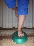
to alternate sitting and standing. If you must stand for a long time, try putting one foot on a low stool.
If you smoke, stop. Smoking decreases blood flow to muscles and disks in and around your spine. Injuries take longer to heal in people who smoke.
Contact your healthcare provider right away if:
You have numbness or tingling in the inner part of your thighs.
You have new or increasing weakness in your legs.
Call your provider for a follow-up appointment if:
The pain is not getting better.
You have new symptoms.

How can I help prevent sciatica?
If you have had back pain and sciatica, you are likely to get it again. To help keep from having it again:
Lose weight if you are overweight.
Do regular aerobic exercise to keep your back and abdominal muscles in shape (this can be as simple as walking),
Do stretching exercises before participating in activities.
Learn to lift properly. Bend your knees and hips and keep your back straight when you lift a heavy object.
Practice good posture. Stand with your head up, shoulders straight, chest forward, weight balanced evenly on both feet, and pelvis tucked in.

https://www.facebook.com/PhysiotherapyCzech

Σπονδυλολίσθιση


Προσοχή: ανοίγει σε νέο παράθυρο. 
Τι είναι η Σπονδυλολίσθηση ; 

Η σπονδυλική μας στήλη αποτελείται από πολλά οστά , τους σπόνδυλος ( βλέπε ανατομία σπονδυλικής στήλης). Κάθε σπόνδυλος συνδέεται με τον άλλον με τις ζυγοαποφυσιακές αρθρώσεις και τον μεσοσπονδύλιο δίσκο. Αυτές οι αρθρώσεις είναι με τέτοιο τρόπο δημιουργημένες έτσι ώστε να φέρουν το βάρος του σώματος και ταυτόχρονα να επιτρέπουν κίνηση. Η σπονδυλολίσθηση αναφέρεται στο « γλίστρημα» ( ολίσθηση) ενός σπονδύλου πάνω στον άλλο ( εικόνα 1 ). Αυτό μερικές φορές συμβαίνει λόγω οστικών ανωμαλιών οι οποίες αναπτύσσονται στην παιδική ηλικία και το παρατηρούμε περισσότερο σε ηλικίες από 9 μέχρι 14 ετών.  Η οστική ανωμαλία μπορεί να είναι η απουσία ενός μέρους του οστού το οποίο κρατά τους σπόνδυλους ευθύγραμμους. Αν και ασυνήθιστο η σπονδυλολίσθηση οφείλεται σε κατάγματα κόπωσης.  
Εικόνα 1-σπονδυλολίσθηση

Κάτι τέτοιο μπορεί να συμβεί κατά την διάρκεια αθλητικής δραστηριότητας η οποία περιλαμβάνει επαναλαμβανόμενη έκταση της σπονδυλικής στήλης. Η σπονδυλολίσθηση βαθμολογείται ανάλογα με το βαθμό μετατόπισης του σπονδύλου σε σχέση με το υποκείμενο του.  Έτσι έχουμε σπονδυλολίσθηση πρώτου βαθμού όπου ο σπόνδυλος μετατοπίζεται λιγότερο με 25% , δευτέρου βαθμού όπου η μετατόπιση είναι 50 % , τρίτου βαθμού 75% και τετάρτου όπου η μετατόπιση είναι μεγαλύτερη από 75% ( εικόνα 2).
        
                            
Εικόνα 2-βαθμοί σπονδυλολίσθησης

Συμπτώματα της σπονδυλολίσθησης 

Οι ασθενείς με σπονδυλολίσθηση συνήθως πονούν κατά την διάρκεια δραστηριοτήτων οι οποίες τείνουν να ευθυγραμμιστούν ή να εκτείνουν της σπονδυλική στήλη , ειδικά αν  αυτές οι δραστηριότητες είναι επαναλαμβανόμενες , υπέρμετρες ή εκτελούνται για μεγάλο χρονικό διάστημα. Αντίστροφα δραστηριότητες οι οποίες τείνουν να κάψουν την σπονδυλική στήλη ( π.χ. το σκύψιμο για να δέσουμε τα κορδόνια μας) μειώνουμε τα συμπτώματα. Είναι σημαντικό να αναφερθεί ότι ασθενείς με σπονδυλολίσθιση πρώτου βαθμού μπορεί να μην βιώσουν ποτέ κανένα συμπτώματα. Αν η σπονδυλολίσθιση είναι από δευτέρου βαθμού και πάνω τότε υπάρχει πόνος στην μέση με η χωρίς αντανακλόμενο πόνο στο πόδι.

Διάγνωση σπονδυλολίσθισης 

Ακτινολογικός έλεγχος συνήθως είναι απαραίτητος για την διάγνωση της σπονδυλολίσθησης ( εικόνα 3 ). Η λήψη της ακτινογραφίας είναι προτιμότερο να γίνει σε θέση πρόκληση των συμπτωμάτων.
     
                                              
Εικόνα 3- ακτινολογική εικόνα της σπονδυλολίσθησης

Θεραπεία σπονδυλολίσθησης 

Η θεραπεία της σπονδυλολίσθησης καθορίζεται από την σταθερότητα των συμπτωμάτων και τον βαθμό του τραυματισμού. Τυπικά η θεραπεία περιλαμβάνει ξεκούραση και αποφυγή των δραστηριοτήτων που αυξάνουν τα συμπτώματα σε συνδυασμό με  άσκηση ενδυνάμωσης τρίτου ή τέταρτου βαθμού πρέπει να αποφύγουν αθλήματα επαφής και υψηλών ταχυτήτων. Δραστηριότητες όπως το κολύμπι , ποδηλασία, κωπηλασία , η γυμναστική Pilate’s αποτελούν καλύτερους και ασφαλέστερους τρόπους εκγύμνασης.

Παράγοντες ανάπτυξης σπονδυλολίσθησης 

Υπάρχουν αρκετοί παράγοντες ανάπτυξης σπονδυλολίσθησης. Όλα πρέπει να αξιολογηθούν και να διορθωθούν από τον φυσικοθεραπευτή. Μερικοί από αυτούς είναι :

  • Λάθος θέση / στάση κορμού
  • Αρθρική αστάθεια
  • Αδυναμία των μυών
  • δυσκαμψία των μυών του κορμού
  • καθιστική ζωή
  • αρθρική δυσκαμψία

Φυσικοθεραπεία για σπονδυλολίσθηση 


Η φυσικοθεραπεία για την σπονδυλολίσθηση είναι ζωτικής σημασία τόσο στην μείωση των συμπτωμάτων όσο και στην ασφαλή επιστροφή στις προηγούμενες δραστηριότητες του ασθενή. Η φυσικοθεραπεία για την σπονδυλολίσθηση περιλαμβάνει :
 

Πηγη  http://www.physio-aid.gr/

ΑΝΑΤΟΜΙΑ ΤΗΣ ΣΠΟΝΔΥΛΙΚΗΣ ΣΤΗΛΗΣ

ΑΝΑΤΟΜΙΑ ΤΗΣ ΣΠΟΝΔΥΛΙΚΗΣ ΣΤΗΛΗΣ
ΟΡΙΣΜΟΣ

Με την αυστηρή (ανατομική) έννοια του όρου, η σπονδυλική στήλη (spine ή vertebral column) είναι ένα σύνολο από οστά (κόκκαλα), κάθε ένα από τα οποία ονομάζεται σπόνδυλος (vertebra). 
Στην πράξη όμως, με τον όρο αυτό, εννοούμε και το εσωτερικό της σπονδυλικής στήλης (δηλ. το νωτιαίο μυελό με την αρχή των νεύρων) αλλά και το εξωτερικό της μέρος (δηλ. τους μύες και συνδέσμους). 

Ένας οσφυϊκός σπόνδυλος

Εικόνα 1 : Ένας οσφυϊκός σπόνδυλος. Στο δεξιό τμήμα της εικόνας φαίνεται η εντομή (σαν άνοιγμα) από όπου εξέρχεται το αντίστοιχο νεύρο. 


Αριθμός και σχήμα των σπονδύλων 

Οι σπόνδυλοι είναι 33. Σε γενικές γραμμές οι σπόνδυλοι έχουν ένα πρόσθιο τμήμα που είναι συμπαγές και λέγεται σώμα (vertebral body) και ένα οπίσθιο τμήμα που λέγεται πέταλο (lamina). Η αφαίρεση του πετάλου είναι η χειρουργική επέμβαση που ονομάζεται πεταλεκτομή. Το πέταλο έχει πολλές προεξοχές (σαν αγκάθια) – κάθε μια λέγεται απόφυση (process). Για παράδειγμα, οι ακανθώδεις αποφύσεις (spinal processes) είναι οι «κόμποι» που ψηλαφάμε στην πλάτη μας, από τον αυχένα μέχρι τη μέση. 
Ανάμεσα στο σώμα και το πέταλο του σπονδύλου, υπάρχει ένα κενό (μια τρύπα). Καθώς ενώνονται οι σπόνδυλοι, ο ένας πάνω στον άλλον σχηματίζεται ένας σωλήνας από αυτά τα «κενά», που έχει ανοίγματα στο πλάι. Ο σωλήνας αυτός λέγεται σπονδυλικός σωλήνας (spinal canal) και μέσα σε αυτόν βρίσκεται προστατευμένος ο νωτιαίος μυελός (spinal cord). Από τα ανοίγματα στο πλάι βγαίνουν τα νεύρα που πηγαίνουν στα χέρια και στα πόδια. 

Ένας οσφυϊκός σπόνδυλος με τα αντίστοιχα νεύρα

1. ΑΥΧΕΝΙΚΗ ΜΟΙΡΑ ή αυχένας (γράφεται και Α.Μ.Σ.Σ. δηλ. αυχενική μοίρα της σπονδυλικής στήλης) από τη βάση του κεφαλιού μέχρι τον προέχοντα σπόνδυλο δηλ. το οστό που ψηλαφάμε στο πίσω μέρος του λαιμού όταν σκύβουμε το κεφάλι μας. Περιλαμβάνει 7 σπονδύλους.

2. ΘΩΡΑΚΙΚΗ ΜΟΙΡΑ (γράφεται και Θ.Μ.Σ.Σ. δηλ. θωρακική μοίρα της σπονδυλικής στήλης) από τον προέχοντα σπόνδυλο μέχρι την αρχή της μέσης. Περιλαμβάνει 12 σπονδύλους.

3. ΟΣΦΥΙΚΗ ΜΟΙΡΑ ή μέση (γράφεται και Ο.Μ.Σ.Σ. δηλ. οσφυϊκή μοίρα της σπονδυλικής στήλης) από το τέλος της θωρακικής μοίρας μέχρι την αρχή του ιερού οστού. Περιλαμβάνει 5 σπονδύλους.

4. ΙΕΡΗ ΜΟΙΡΑ . Περιλαμβάνει 5 σπονδύλους ενωμένους μεταξύ τους, που αποτελούν το ιερό οστό.

5. ΚΟΚΚΥΓΙΚΗ ΜΟΙΡΑ. Περιλαμβάνει 4 πολύ μικρούς σπονδύλους, ενωμένους μεταξύ τους, που αποτελούν τον κόκκυγα.

ΔΥΟ ΓΕΙΤΟΝΙΚΟΙ ΣΠΟΝΔΥΛΟΙ (ΦΩΤΟ) ενώνονται μεταξύ τους με το μεσοσπονδύλιο δίσκο, τις αρθρώσεις και τους συνδέσμους :

1. Ο ΜΕΣΟΣΠΟΝΔΥΛΙΟΣ ΔΙΣΚΟΣ (intervertebral disc) είναι ένα ελαστικό «μαξιλαράκι» που ενώνει τα σώματα των σπονδύλων και απορροφά τους κραδασμούς. Η φθορά του δίσκου, είτε απότομα, είτε συνηθέστερα σε χρόνια βάση, προκαλεί πίεση στα νεύρα και το νωτιαίο μυελό (βλ. αυχενική δισκοκήλη, οσφυϊκή δισκοκήλη). Ο μεσοσπονδύλιος δίσκος αποτελείται από ένα περίβλημα (ινώδης δακτύλιος) και το εσωτερικό του (πηκτοειδής πυρήνας). Ο πυρήνας είναι μαλακός σαν τσίχλα και έχει μεγάλη ποσότητα νερού.

2. Η ΑΡΘΡΩΣΗ (facet joint) σχηματίζεται από ένα τμήμα του πάνω σπονδύλου και άλλο ένα του κάτω σπονδύλου. Για κάθε ζεύγος σπονδύλων, υπάρχουν δύο αρθρώσεις, μια δεξιά και μια αριστερά

3. ΟΙ ΣΥΝΔΕΣΜΟΙ (ligaments) είναι «λαστιχάκια» που συνδέουν τα πέταλα δύο γειτονικών σπονδύλων.

πηγη http://www.neurocenter.gr/

Σάββατο 15 Φεβρουαρίου 2014

Διαστρέμματα ποδοκνημικής, οξύ & χρόνιο διάστρεμμα – Θεραπευτικές επιλογές.


Οι κακώσεις συνδέσμων στην έξω επιφάνεια του αστραγάλου και  ο τραυματισμός των εξωτερικών συνδέσμων είναι από τους ποιο συχνούς τραυματισμούς στον αθλητικό πληθυσμό  Εκτιμάται ότι αυτοί οι τραυματισμοί αποτελούν  περίπου το 25% από το σύνολο των αθλητικών τραυματισμών. 

Οξύς τραυματισμός των συνδέσμων

Η ποιο ευάλωτοι εξωτερικοί πλάγιοι σύνδεσμοι είναι ο πρόσθιος αστραγαλοπερονικός σύνδεσμος,  συχνά ακολουθείτε  μια συνδυασμένη ρήξη αυτού του συνδέσμου και του πτερνοπερονικού συνδέσμου  Άλλοι τραυματισμοί όπως ο τραυματισμός του έσω δελτοειδούς συνδέσμου είναι λιγότεροι συχνοί, αποτελώντας λιγότερο από το 10% από όλους τους τραυματισμούς. Ο μηχανισμός του τραυματισμού για τους έξω πλάγιους συνδέσμους συμβαίνει ποιο συχνά σε μία θέση πελματιαίας κάμψης ( Plantar flexion)  και ανάσπαση του έσω χείλους (Inversion). 


Η γενικές αρχές θεραπείας και η διαχείρηση ενός οξύ μιας πρόσφατης ρίξης συνδέσμου περιλαμβάνει:

  • Ανάπαυση
  • Ακινητοποίηση
  •  Πάγο και ανάροπη θέση.


Οι τελευταίες επιστημονικές μελέτες  αποδεικνύουν πως η έγκαιρη διάγνωση, η σωστή θεραπευτική επιλογή και το κατάλληλο πρόγραμμα αποκατάστασης είναι πολύ σημαντικά για την πρόληψη της χρόνιας πλευρικής συνδεσμικής αστάθειας του αστραγάλου ( χρόνιο διάστρεμμα . Οι πρόσφατες έρευνες αποκαλύπτουν και αποδεικνύουν πως το 75% περίπου όλων των συνδεσμικών τραυματισμών στον αστράγαλο  επανεμφανίζεται και μεταπίπτει σε μια χρόνια φάση δημιουργώντας χρόνια αστάθεια στην άρθρωση με τα ανάλογα συμπτώματα και λειτουργικούς περιορισμούς στον αθλητή ή τον ασθενή. Επίσης έχει αποδειχθεί από πολλές μελέτες ότι η χρόνια εξωτερική αστάθεια της αστραγακοπτερνικής άρθρωσης μπορεί να αναπτυχθεί περίπου στο 10% με 30% σε όλους τους ασθενείς που διατηρούν ένα οξύ τραυματισμό συνδέσμου. 

Πρόληψη

Ο καλύτερος τρόπος για να θεραπεύσουμε τον  τραυματισμό των συνδέσμων είναι να εμποδίσουμε να συμβεί αυτό, αν και αυτό πρακτικά είναι αδύνατον. Παρόλα αυτά η πρόληψη μπορεί να παίξει σημαντικό ρόλο. Η Πρόληψη μπορεί να γίνει είτε με προπόνηση συντονισμού χρησιμοποιώντας ειδικές ασκήσεις πάνω σε  σανίδες ισορροπίας και ειδικά μαλακά δάπεδα ( Stifness)  ή με εξωτερική υποστήριξη της άρθρωσης χρησιμοποιώντας ειδικές ταινίες υποστήριξης (Kinesio Tape), νάρθηκες κ.τ.λ.

πηγη http://rehabilitimifizioterapeutik.blogspot.gr/